Protein-like nanoparticles sort themselves inside growing crystals, enabling controlled release

The tiny bones in your fingers withstand countless taps and swipes thanks to a precise blend of materials. Flexible collagen fibers form the framework, reinforced by hard calcium phosphate hydroxyapatite crystals. This is just one of countless examples in which living organisms weave organic materials directly into inorganic crystals with exquisite precision. In a recent study published in Nature Communications, scientists attempted to recreate such precise spatial arrangements in biomimetic composite materials.
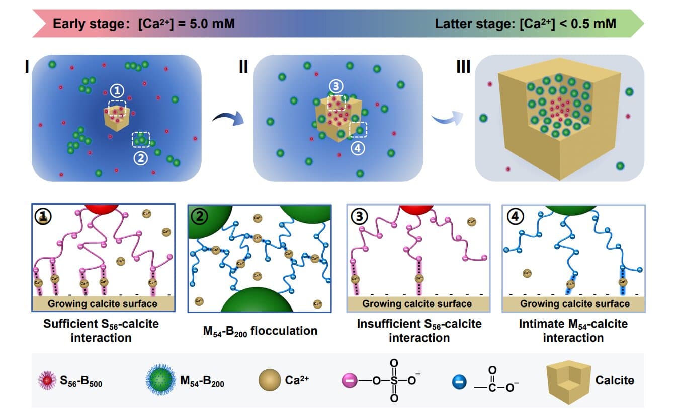
The researchers made two types of tiny diblock copolymer nanoparticles designed to mimic pseudo-proteins. The first consisted of solid spheres about 100 nanometers big, with a poly(benzyl methacrylate) core and a shell of sulfate-containing chains tagged with a red fluorescent dye. The second consisted of hollow, bubble-like particles about 300 nanometers across. They had the same polymer core but a carboxylate-rich outer shell tagged with a green fluorescent dye.
These tiny polymer nanoparticles, engineered with different sizes and compositions, much like protein molecules, sorted themselves naturally as growing calcite crystals trapped them. Instead of mixing randomly, the two types ended up in separate regions of the crystal, creating an artificial biomineral with a distinctly organized structure—all driven by differences in the nanoparticles' surface chemistry.
Mimicking nature's materials lab
In natural biominerals such as bones, teeth and shells, hard inorganic minerals are combined with organic molecules such as peptides, forming highly organized hybrid structures. Their durability and unique properties have long prompted scientists to try to recreate them by growing synthetic crystals with similar organic-inorganic architectures. Studies have managed to trap a single type of nanoparticle inside a growing crystal, but natural biomineralization involves many different components. What they lacked were clear rules for making multiple particle types sort themselves into specific regions as a crystal grows.
At such tiny scales, existing analytical techniques could not reliably untangle the overlapping interactions between multiple components, let alone track them individually.
To overcome these issues, researchers synthesized nanoparticles with precisely controlled surface chemistries and sizes, using them as tunable stand-ins for natural biomolecules. This provided a new experimental model for exploring the factors that govern nanoparticle partitioning and segregation during crystal growth.
The two synthetic protein stand-ins were built using a precise chemical technique called RAFT polymerization combined with polymerization-induced self-assembly (PISA). To make them visible under advanced microscopes, the smaller spheres (S56-B500) were dyed red, while the hollow, bubble-like vesicles (M54-B200) were dyed green. They grew calcite crystals over the course of a day, placing glass slides at the bottom of a liquid dish containing a mixture of both nanoparticle types. As calcium slowly crystallized onto the slides, it trapped the nanoparticles inside the forming mineral.

Controlled synthesis of diblock copolymer spheres or vesicles via RAFT-mediated PISA. Credit: Nature Communications (2026). DOI: 10.1038/s41467-026-75685-3 
In situ monitoring of the self-sorting occlusion of S56 B500 spheres and M54 B200 vesicles during calcite crystal growth. Credit: Nature Communications (2026). DOI: 10.1038/s41467-026-75685-3
Using a specialized laser microscope, they scanned the crystals, tracking the red and green dyes to build a 3D map of the particle locations.
They discovered that different populations of synthetic nanoparticles spontaneously organized and sorted themselves into separate layers inside a single growing crystal. Sulfate-coated particles were captured early and trapped in the crystal core, while carboxylate-coated particles joined later, forming a distinct layer near the surface.
The sorting was determined strictly by the surface chemistry of the nanoparticle coating, not by their size or shape. As calcium ions were consumed during crystal growth, changing chemical conditions shifted which particles could enter next.
Calcite is pH-sensitive and dissolves from the outside when exposed to acid, which meant the crystal design came with a built-in release schedule. When placed in mild acid at pH 4, they observed that the outer green vesicles were released first, and the red core spheres escaped only after the crystal's center dissolved.
The researchers noted that this level of control over nanoscale organization allows multifunctional composite crystals to be built with a clear plan rather than trial and error. The findings also deepen our understanding of how nature forms complex mineral structures, knowledge that might come in handy when engineering precise drug delivery systems.
Written for you by our author Sanjukta Mondal, edited by Robert Egan—this article is the result of careful human work. We rely on readers like you to keep independent science journalism alive. If this reporting matters to you, please consider a donation (especially monthly). You'll get an ad-free account as a thank-you.
Publication detailsWenting Chen et al, Interfacial chemistry governs nanoparticle self-sorting during biomimetic crystallization, Nature Communications (2026). DOI: 10.1038/s41467-026-75685-3
© 2026 Science X Network
Citation: Protein-like nanoparticles sort themselves inside growing crystals, enabling controlled release (2026, August 13) retrieved 13 August 2026 from https://phys.org/news/2026-08-protein-nanoparticles-crystals-enabling.html
This document is subject to copyright. Apart from any fair dealing for the purpose of private study or research, no part may be reproduced without the written permission. The content is provided for information purposes only.
Reporting from Phys.org contributed to this story.